Categories
Feature Health & Medicine Researcher news Slideshows Student researchers

NCI grant funds SMU research into cancer-causing viruses that hide from the immune system

Genes common to both the human T-cell leukemia virus and high-risk human papillomaviruses activate survival mechanisms in cancer cells. An SMU lab, with National Cancer Institute funding, is hunting ways to inhibit those genes to halt the development of cancer.

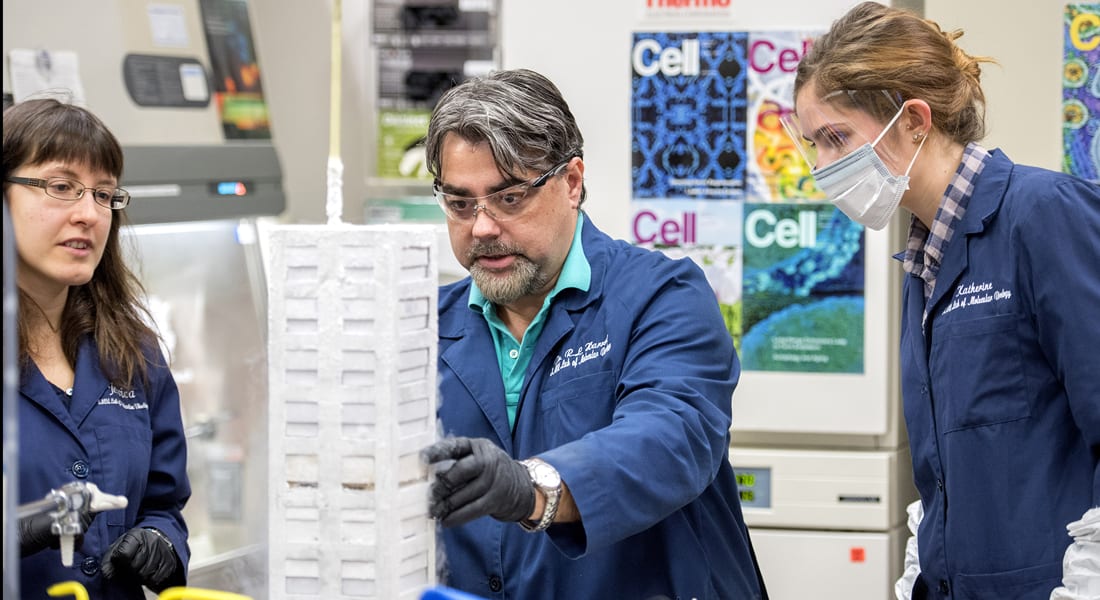
SMU virologist and cancer researcher Robert L. Harrod has been awarded a $436,500 grant from the National Cancer Institute to further his lab’s research into how certain viruses cause cancers in humans.

Under two previous NCI grants, Harrod’s lab discovered that the human T-cell leukemia virus type-1, HTLV-1, and high-risk subtype human papillomaviruses, HPVs, share a common mechanism that plays a key role in allowing cancers to develop.

Now the lab will search for the biological mechanism — a molecular target — to intervene to block establishment and progression of virus-induced cancers. The hope is to ultimately develop a chemotherapy drug to block the growth of those tumor cells in patients.

“The general theme of our lab is understanding the key molecular events involved in how the viruses allow cancer to develop,” said Harrod, an associate professor in SMU’s Department of Biological Sciences whose research focuses on understanding the molecular basis of viral initiation of cancer formation.

While HTLV-1 and HPV are unrelated transforming viruses and lead to very different types of cancers, they’ve evolved a similar mechanism to cooperate with genes that cause cancer in different cell types. The lab discovered that the two viruses tap a common protein that cooperates with cellular genes to help the viruses hide from the immune system.

That common protein, the p30 protein of HTLV-1, binds to a different protein in the cell, p53, which normally has the job of suppressing cancerous growth or tumor development. Instead, however, p30 manages to subvert p53’s tumor suppressor functions, which in turn activates pro-survival pathways for the virus.

From there, the virus can hide inside the infected cell for two to three decades while evading host immune-surveillance pathways. As the cell divides, the virus divides and replicates. Then ultimately the deregulation of gene expression by viral encoded products causes cancer to develop.

“They are essentially using a similar mechanism, p30, to deregulate those pathways from their normal tumor-suppressing function,” Harrod said.

Tumor suppression, DNA damage-repair pathways, begin to fail with age
About 15 percent to 20 percent of all cancers are virus related. Worldwide, about 10 million people are infected with HTLV-1 and, as with other viral-induced cancers, about 3 percent to 5 percent of those infected go on to develop malignant disease.

Cancer is often associated with the process of normal aging, because our tumor suppression and DNA damage-repair pathways begin to break down and fail, explained Harrod. Our pathways don’t as easily repair genetic mutations, which makes us more susceptible to cancers like adult T-cell leukemia and HPV-associated cervical cancers or head-and-neck carcinomas, he said.

The human T-cell leukemia virus is transmitted through blood and body fluid contact, usually infecting infants and children via breastfeeding from their mother. A tropical infectious disease, it’s endemic to Southeast Asia, primarily Japan, Taiwan, China and Malaysia, as well as certain regions in the Middle East, Northern Africa and islands of the Caribbean. In the United States, Hawaii and Florida have the highest incidence of adult T-cell leukemia. HTLV-1 is highly resistant to most modern anticancer therapies, including radiotherapy and bone marrow or matching donor stem cell transplants. The life expectancy of patients with acute or lymphoma-stage disease is about six months to two years after diagnosis.

In the case of HPV, certain high-risk sub-types aren’t inhibited by today’s available HPV vaccines. It’s considered the high-risk HPVs are sexually transmitted through direct contact with the tissues of the virus-producing papillomas or warts. High-risk HPVs can also cause cervical cancers and head and neck carcinomas, many of which are associated with poor clinical outcomes and have high mortality rates.

How do viruses cause cancer?
For both HTLV-1 and HPV, the virus itself does not cause cancer to develop.

“It’s cooperating with oncogenes — cellular genes that become deregulated and have the potential to cause cancer,” Harrod said. “The role of these viruses, it seems, is to induce the proliferation of the cell affected with cancer. We’re trying to understand some of the molecular events that are associated with these cancers. ”

The lab’s three-year NCI grant runs through 2019. Harrod’s two previous grants awarded by the National Institutes of Health were also three-year-grants, for $435,000 and $162,000. Each one has targeted HTLV-1 and the p30 protein.

The lab’s first NCI grant came after the researchers provided the first demonstration that p30 could cooperate with cellular oncogenes, which have the potential to cause cancer, to cause deregulated cell growth leading to normal cells transforming into cancer cells. That original discovery was reported in 2005 in the article “A human T-cell lymphotropic virus type 1 enhancer of Myc transforming potential stabilizes Myc-TIP60 transcriptional interactions,” in the high-profile journal Molecular and Cellular Biology.

“We find that the p30 protein is involved in maintaining the latency of these viruses. These viruses have to persist in the body for 20 to 40 years before a person develops disease. To do that they have to hide from the immune response,” Harrod explained. “So p30 plays a role in silencing the viral genome so that the affected cells can hide, but at the same time it induces replication of the affected cells. So when the cell divides, the virus divides. We call that pro-viral replication.”

The term “latency maintenance factor” in reference to p30 originated with Harrod’s lab and has gained traction in the HTLV-1 field.

Under the lab’s second NCI grant, the researchers figured out how to block pro-survival pathways to kill tumor cells.

In the current grant proposal, Harrod’s lab demonstrated that by inhibiting specific downstream targets of p53 — essentially blocking pathways regulated by the p53 protein — they could cause infected tumor cells to collapse on themselves and undergo cell death.

“We do that independent of chemotherapy,” Harrod said. “So that was a big find for us.”

Goal is to eliminate cancer cells by inhibiting pathway
Each grant project builds upon the one before it, and the third grant extends the work, to now include high-risk HPVs.

“Now that we’ve shown we can block one or two of these factors to cause cell death, we’re starting to get an eye really on how we can inhibit these cancer cells and what potentially down the road may lead to a therapeutic,” Harrod said. “That’s the ultimate goal.”

One of the biggest challenges will be to inhibit the pathways in the tumor cells without targeting normal cells, he said. The lab’s recent findings indicate the researchers may soon be within reach of identifying a new strategy to eliminate cancer cells by inhibiting pathways key to their survival.

Harrod’s lab collaborates on the research with: Lawrence Banks, Tumor Virology Group Leader, International Centre for Genetic Engineering and Biotechnology, Trieste, Italy; Brenda Hernandez, Associate Director, Hawaii Tumor Registry, University of Hawaii Cancer Center, Honolulu; and Patrick Green, Director, Center for Retrovirus Research, The Ohio State University. — Margaret Allen, SMU
Categories
Culture, Society & Family Feature Fossils & Ruins Researcher news Technology

Evidence of first chief indicates Pacific islanders invented a new society on city they built of coral and basalt

New analysis of chief’s tomb suggests island’s monumental structures are earliest evidence of chiefdom in Pacific — yielding new keys to how societies emerge and evolve

New dating on the stone buildings of Nan Madol suggests the ancient coral reef capital in the Pacific Ocean was the earliest among the islands to be ruled by a single chief.

The discovery makes Nan Madol a key locale for studying how ancient human societies evolved from simple societies to more complex societies, said archaeologist Mark D. McCoy, Southern Methodist University, Dallas. McCoy led the discovery team.

The finding was uncovered as part of a National Geographic expedition to study the monumental tomb said to belong to the first chief of the island of Pohnpei.

McCoy deployed uranium series dating to determine that when the tomb was built it was one-of-a-kind, making it the first monumental scaled burial site on the remote islands of the Pacific.

The discovery enables archaeologists to study more precisely how societies transform to more and more complex and hierarchical systems, said McCoy, an expert in landscape archaeology and monumental architecture and ideology in the Pacific Islands.

“The kind of society that we live in today, it wasn’t born last year, or even 100 years ago,” McCoy said. “It has its roots in a pre-modern era like Nan Madol where you have a king or chief. These islanders invented a new kind of society — that is a socially creative achievement. The idea of chiefs, someone in charge, is not a new thing, but it’s an extremely important precursor. We know tribes and bands predate chiefdoms and states. But it’s not a straight line. By looking at these intermediate stages we get insight into that social phenomenon.”

The analysis is the first time uranium-thorium series dating, which is significantly more precise than previously used radiocarbon dating, was deployed to calculate the age of the stone buildings that make up the famous site of Nan Madol (pronounced Nehn Muh-DOLL) – the former capital of the island of Pohnpei.

“The thing that makes this case special is Nan Madol happened in isolation, it happened very recently, and we have multiple lines of evidence, including oral histories to support the analysis,” McCoy said. ”And because it’s an island we can be much more specific about the natural resources, the population, all the things that are more difficult when people are on a continent and all connected. So we can understand it with a lot more precision.”

Nan Madol, which UNESCO this year named a World Heritage Site, was previously dated as being established in A.D. 1300. McCoy’s team narrowed that to just a 20-year window more than 100 years earlier, from 1180 to 1200.

The finding pushes back even earlier the establishment of the powerful dynasty of Saudeleur chiefs who asserted authority over the island society for more than 1,000 years.

First chief was buried in Pohnpei tomb by A.D. 1200
An ancient city built atop a coral reef, Nan Madol has been uninhabited for centuries now. Located in the northwestern Pacific on the remote island of Pohnpei, it’s accessible via a 10-hour flight from Hawaii interspersed with short hops from atoll to atoll, including a stop at a U.S. military installation. Nan Madol is the largest archaeological site in Micronesia, a group of islands in the Caroline Archipelago of Oceania.

Uranium dating indicates that by 1180, massive stones were being transported from a volcanic plug on the opposite side of the island for construction of the tomb. And by 1200, the burial vault had its first internment, the island’s chief.

Construction of monumental buildings followed over the next several centuries on other islands not in the Saudeleur Dynasty across Oceania.

McCoy, an associate professor in the SMU Department of Anthropology, and his team reported their discovery in the journal Quaternary Research in “Earliest direct evidence of monument building at the archaeological site of Nan Madol identified using 230Th/U coral dating and geochemical sourcing of megalithic architectural stone.”

Co-authors include Helen A. Alderson, University of Cambridge, U.K., Richard Hemi, University of Otago, New Zealand, Hai Cheng, Xi’an Jiaotong University, China, and R. Lawrence Edwards, University of Minnesota.

An inactive volcano that hasn’t erupted in at least one million years, Pohnpei Island is much larger than its neighboring atolls at 128 square miles (334 square kilometers), making it about the physical size of Columbia, S.C.

Now part of the 607-island nation of the Federated States of Micronesia, Pohnpei Island and its nearby atolls have a population of 34,000.

Pohnpei monument indicates invention of a new kind of society
How Nan Madol was built remains an engineering mystery, much like Egypt’s Pyramids.

“It’s a fair comparison to the Pyramids, because the construction, like the Pyramids, didn’t help anyone — it didn’t help society be fairer, or to grow crops or to provide any social good. It’s just a really big place to put a dead person,” McCoy said.

It’s important to document such things, he said, because this architectural wonder indicates that independently of Egypt, another group of people put effort into building a monument.

“And we think that’s associated with the invention of a new kind of society, a new kind of chiefdom that ruled the entire island,” McCoy said.

Unlike Egypt and the Pyramids however, Nan Madol was invented much more recently in the big story of human prehistory, he said.

“At A.D. 1200 there are universities in Europe. The Romans had come and gone. The Egyptians had come and gone,” he said. “But when you’re looking at Pohnpei, it’s very recent, so we still have the oral histories of the descendants of the people who built Nan Madol. There’s evidence that you just don’t have elsewhere.”

Monumental city built of coral and stone
Pohnpei was originally settled in A.D. 1 by islanders from the Solomon or Vanuatu island groups. According to local oral history, the Saudeleur Dynasty is estimated to have begun its rule around 1160 by counting back generations from the modern day.

To build the tomb and other structures, naturally formed boulders of basalt, each weighing tons, were somehow transported far from existing quarries on the other side of the island to a lagoon overgrown with mangrove and stretching across 205 acres (83 hectares).

The basalt blocks formed when hot lava cooled and adopted the shape of long, column-shaped boulders and cobbles. Formed from 1 million to 8 million years ago, they came from a number of possible quarry locations on the island.

The city’s stone structures were built atop 98 shallow artificial coral reef islets, each one built by the Saudeleur people. The structures were constructed about three feet above waterline by laying down framing stones, filling the void between them with crushed coral, then laying up double parallel walls and again filling the gap between with crushed coral. The islets are separated by tidal canals and protected from the ocean by 12 sea walls, making Nan Madol what many consider the Venice of the Pacific.

“The structures are very cleverly built,” said McCoy. “We think of coral as precious, but for the architects of Nan Madol it was a building material. They were on a little island surrounded by huge amounts of coral reef that grows really quickly in this environment, so they could paddle out at low tide and mine the coral by smashing some off and breaking it up into rubble.”

The largest and most elaborate architecture in the city is the tomb of the first Saudeleur, measuring 262 feet by 196 feet (80 meters by 60 meters), basically the size of a football field. It is more than 26 feet (8 meters) tall, with exterior walls about six feet to 10 feet (1.8 to 3 meters) thick. A maze of walls and interior walkways, it includes an underground crypt capped with basalt.

“The architecture is meant to be extremely impressive, and it is,” McCoy said. “The structures were built to last — this is one of the rainiest places on earth, so it can be muddy and slippery and wet, but these islets on the coral reef are very stable.”

Portable X-ray technology provides clue to source of megalithic stones
McCoy and his team used portable X-ray fluorescence (XRF) to geochemically match the columnar-shaped basalt stones to natural sources on the island. The uranium-thorium technique calculates a date based on characteristics of the radioactive isotope thorium-230 and its radioactive parent uranium-234.

That enabled them to determine the construction chronology of a tomb that oral histories identify as the resting place of the first chief to rule the entire island.

“We used an X-ray gun, which looks like a 1950s-styled ray gun,” McCoy said. “It allows you — at a distance and without destroying the thing you’re interested in — to bounce X-rays off it and work out what the chemistry is. The mobile technology has gotten much more affordable, making this kind of study feasible.”

Using uranium series dating on coral emerged in the last decade. Accuracy — superior to radiocarbon — is plus or minus a few years of when the coral died. A very good radiocarbon date only will get within 100 years.

“That’s a monumental shift in terms of the precision with which we talk about things,” McCoy said. “If Nan Madol had not been made of the kind of stone we could source, if the architects hadn’t chosen to use coral, we wouldn’t have been able to get this date. So it’s a happy coincidence that the evidence at the site came together.”

McCoy suggests that future research look at finding the cause for this major turning point on Pohnpei, and what sparked this new hierarchy of rule and monumental building in this society. — Margaret Allen, SMU

Categories
Feature Health & Medicine Mind & Brain Researcher news Videos

Researchers test blood flow in athletes’ brains to find markers that diagnose concussions

Diagnosing concussions is difficult because it typically rests on subjective symptoms such as forgetfulness, wobbly gait and disorientation or loss of consciousness. A new study of college athletes investigates objective indicators using Doppler ultrasound to measure brain blood flow and blood vessel function.

A hard hit to the head typically prompts physicians to look for signs of a concussion based on symptoms such as forgetfulness, wobbly gait and disorientation.

But symptoms such as those are subjective. And youth who are anxious to get back to their sport can sometimes hide the signs in order to brush off adult concerns, says physiologist Sushmita Purkayastha, Southern Methodist University, Dallas.

Now a new study funded by the Texas Institute for Brain Injury and Repair at U.T. Southwestern Medical Center, Dallas aims to find noninvasive objective indicators to diagnose whether an athlete has suffered a concussion. Using transcranial Doppler ultrasound, the study will probe the brains of college athletes to measure blood vessel function in the brain, looking for tell-tale signs related to blood flow that help diagnose concussion, said Purkayastha, a researcher on the new study.

“We know this is an understudied area. With other health problems, when the doctor suspects diabetes or hypertension, they don’t guess, they run objective tests to confirm the diagnosis. But that’s not the case with concussion — yet,” said Purkayastha, whose research expertise is blood flow regulation in the human brain. “That’s why my research focus is to find markers that are objective and not subjective. And this method of monitoring blood flow in the brain with ultrasound is noninvasive, inexpensive and there’s no radiation.”

Purkayastha and others on the research team are working under a one-year, $150,000 pilot research grant from the Texas Institute for Brain Injury and Repair, a UT Southwestern initiative funded by the Texas Legislature to enhance the diagnosis and treatment of brain injuries.

The team will observe 200 male and female college athletes over the next two years. Half the athletes will be students playing a contact-collision sport who have recently suffered a sports-related concussion. The other half, a control group, will be students playing a contact-collision sport who don’t have a concussion. The study draws on athletes from football, soccer, equestrian sports, cheerleading and recreational sports.

The researchers began testing subjects in August. They expect to have results by the Fall of 2017.

“We are very excited at establishing this collaboration between SMU and the Physical Medicine and Rehabilitation Department at UTSW. Our work with Dr. Purkayastha promises to give meaningful insight into the role of cerebral blood flow mechanisms after concussion and will point us in the right direction for improved neurorecovery,” said physician Kathleen Bell, a leading investigator at U.T. Southwestern’s Texas Institute for Brain Injury and Repair and principal investigator on the study. Bell is a nationally recognized leader in rehabilitation medicine and a specialist in neurorehabilitation.

Diagnosing concussions by using objective, non-invasive and inexpensive markers will result in accurate diagnosis and better return-to-play decisions following a concussion, thereby preventing the long-term risk of second-impact syndrome, said Purkayastha, an assistant professor in the Department of Applied Physiology and Wellness of SMU’s Annette Caldwell Simmons School of Education and Human Development.

“Although sports-related concussions are common, the physiology of the injury is poorly understood, and hence there are limited treatments currently available,” she said.

Hemorrhage or blackouts result, for example, if autoregulation malfunctions
While the brain is the most important organ in the body, it has been very understudied, said Purkayastha, a professor in the Simmons School of Education & Human Development. But since blood vessels in the brain behave similarly to those in the rest of the body, it’s possible to measure blood vessel function in the brain by monitoring blood pressure and brain blood flow. Observing those functions could reveal a marker, she said.

In Purkayastha’s lab on the SMU campus, student athletes are being outfitted with two small ultrasound probes, one on each side of their forehead in the temple area, to test blood vessel function. Specifically, the two probes monitor the blood flow through middle cerebral artery, which supplies blood to 75 percent of the brain. The artery traverses the brain, circulating blood to the brain tissues responsible for movement, cognition and decision-making.

Branching from the middle cerebral artery is a network of blood vessels that get smaller and smaller as they get further from the artery, spreading like tree branches through the brain. The smallest vessels — via a different local regulatory mechanism — maintain constant blood flow to the brain, making microadjustments, such as constricting and dilating in the face of constant changes in blood pressure. Adjustments occur as a person’s muscles move, whether standing, sitting, exercising, or even just laughing and experiencing emotion. These continual adjustments in the vessels — called cerebral autoregulation — keep blood flow constant and regular. That prevents problems such as hemorrhaging or passing out from large fluctuations in blood pressure that is either too high or too low.

Researchers suspect concussion diminishes a vessels ability to properly regulate blood flow
In the current study, ultrasound probes on the temples record the vessels’ microadjustments as digital data. That information is processed through a WinDaq data acquisition software and analyzed to examine cerebral autoregulation with spontaneous changes in blood pressure during that period of time.

Unlike at the doctor’s office, when a cuff is used to measure blood pressure at a rate of single measurements during 30 seconds, Purkayastha’s ultrasound monitoring of blood pressure provides continuous blood pressure recording throughout each heartbeat. As sound waves bounce into the artery and send back an echo, they measure the speed of red blood cells and other blood components moving through the artery.

“We collect 10 minutes of very high frequency data points collecting information on beat-to-beat changes in blood pressure and blood flow to the brain for every single heartbeat,” said Purkayastha. “Then we analyze and post-process and examine how well the blood vessels were able to maintain constant blood flow to the brain. We suspect in people with concussion that the autoregulation function isn’t operating properly which leads to impairments in function such as wobbly gait, disorientation or forgetfulness. This is a noninvasive way to see if there’s a flaw in the autoregulation.”

Athletes with confirmed diagnosis of concussions will be tested three times during the course of the study. The first test is three days after a suspected concussion, the second is 21 days afterward, and the third is three months afterward.

“The pilot studies so far look promising and our goal is to better understand the mechanism behind injury and design objective markers detecting concussion,” said Purkayastha.

The Texas Institute for Brain Injury and Repair at U.T. Southwestern Medical Center, a component of the Harold and Annette Simmons Comprehensive Center for Research and Treatment in Brain and Neurological Disorders, is a collaborative initiative involving local and national organizations, including the National Institutes of Health, University of Texas Dallas and its Center for BrainHealth, Children’s Medical Center, Dallas VA Medical Center, and Parkland Health and Hospital System, as well as Texas Health Resources and Texas Health Ben Hogan Sports Medicine. — Margaret Allen, SMU